Equine Fact Sheets
Explore our equine fact sheets, which cover many common health concerns in horses.
Scroll down and click the tabs to learn more.
Equine wormer resistance
What is wormer resistance?
Anthelmintic resistance occurs when a proportion of the parasites picked up from a particular pasture are no longer affected by the chosen worming treatment. A number of factors can contribute to the development of resistance, including underdosing and the frequent, perhaps unnecessary, administration of wormers over time.
All worms in any given population (i.e. inside a horse) are each as genetically different from each other as humans are genetically different from one another. A very few of those worms will have a natural genetic variation that can enable them to survive a dose of anthelmintic, or wormer. This sort of variation exists within all populations of all organisms. This occurs partly because random mutations occur in the genome of an individual organism, and these mutations can be passed to offspring. Throughout the individuals’ lives, their genomes interact with their environments to cause variations in traits. The process of natural selection, as detailed by Charles Darwin’s theory of evolution, states that those living things that are the best adapted to survive in their environments will survive and be the most successful at reproduction, therefore having more offspring, which also have the genetic mutation. Eventually, through natural selection such mutations become the norm.
How does wormer resistance develop?
Using too low a dose of wormer is one of the key ways that helps those slightly, naturally resistant worms to survive. . These then breed with each other to potentially produce a higher proportion of resistant worms and a -smaller population of susceptible worms within the population. Successive doses of the same wormer, or wormers from the same drug class, can lead to an established resistant population.
Frequent, unnecessary worming can also increase the potential for resistance, by actively selecting for worms that are resistant and killing out susceptible worms before they can reach sexual maturity and produce offspring.
Once resistance is present in a worm population, the health, welfare and performance of those horses infested with resistant worms will be compromised. It is also impossible to revert back to a susceptible population.
Reducing and preventing resistance
The key to reducing the likelihood of resistance developing starts with the identification of those horses which need to be treated. This can be achieved by testing individual horses, thereby identifying those animals with a significant worm burden, and then using the correct wormer to treat them – calculating the correct dose and time to worm.
Worm egg counts help to ensure that wormers are only given when they are needed and therefore may reduce the likelihood of resistance developing. This helps your individual horse and the equine community as a whole.
As part of this more targeted approach to worming, industry experts are calling for horse owners to make better use of Worm Egg Counts (WECs). A WEC is a microscopic examination of a dung sample from a horse to detect and count the number of roundworm eggs present. The egg count is expressed as eggs per gram (epg), and in most cases if the count is greater than 200epg then worming should be considered (some foals and horses, especially where there is no previous worming history, may still require treatment where the worm egg count is less than 200 epg; consult your vet for further advice). A WEC therefore helps to identify those horses that have an excessive worm burden and would benefit from a treatment. It also identifies the main species of worms with the exception of tapeworms, and immature and encysted worms.
It is known that approximately 80% of worms are carried by only 20% of horses. The regime of regular worming at fixed intervals, a system that has traditionally been popular on large yards, has provided an effective means of limiting worm burdens, but it is possible that many horses are being wormed unnecessarily, which encourages the development of resistance. With the consensus of expert opinion on the future of worming firmly behind the use of targeted programmes incorporating WECs, there has never been a better time to make worm egg counts a regular part of your worming programme.
Keeping track of treatments and WEC results may seem like an onerous task, however Merial’s online SMART planner reminds you when to test and worm accordingly, helping you to keep track of every horse in your yard if required.
Find out more at http://www.smartworming.co.uk/
Equine faecal samples
Providing a faecal sample for worm egg counts (WEC)
Worm Egg Counts (WEC) are an essential part of your horse’s health care routine. They help monitor and manage your horse’s internal parasite levels, allowing you and your vet to make informed decisions about whether deworming is necessary.
Regular WEC testing helps reduce unnecessary use of wormers, slows the development of resistance in parasites and keeps your horse healthier in the long term.
To ensure you receive accurate and meaningful results, it’s important to collect and submit your sample correctly. Download our guide below for steps on how to get the best outcome.
Equine strangles
The disease
Strangles is a widespread, highly infectious, debilitating disease of horses. It is caused by a bacterium called streptococcus equi. Strangles is considered to be the most commonly diagnosed infectious disease affecting horses of all sizes, breeds, types and ages, although some groups are higher risk than others and may be the worst affected – young horses (<5 years old), sick horses, horses with compromised immune systems, stud farms, racing stables, livery yards and riding school horses. This is due to the way the disease is spread. Direct contact between horses is the most common factor, but spread of the disease is also via contaminated food, drinking vessels or equipment including people’s clothing. The disease can be spread by inhalation but this is less common.
Strangles is not a notifiable disease, but yards with confirmed outbreaks are strongly recommended to implement stringent biosecurity precautions to prevent further spread, and to notify neighbouring premises.
Clinical signs
The severity of clinical signs can vary depending on the age and condition of the horse. Clinical signs are usually seen between 3-7 days after the horse has been in contact with the bacterium, but can take as long as 14 days to show.
Clinical signs include:
- Loss of appetite
- Difficulty in swallowing
- Nasal discharge
- Depression and dullness
- Development of a cough
- Fever
- Swelling of the lymph nodes (glands) under the jaw or lower down the neck approximately a week after the onset of clinical signs.
- Complications seen in a small number of rare cases include:
- Abscesses placing pressure on the airways, causing the horse to suffocate – hence the term ‘Strangles’
- ‘Bastard Strangles’ causes swelling of lymph nodes in other areas of the body such as the brain, lungs and/or intestines. Rupture of these abscesses is usually fatal.
- Purpura haemorrhagica which causes bleeding into the skin, gums and organs such as the lungs and can also prove fatal.
When the abscesses in the lymph nodes burst they discharge a highly infectious, thick creamy-yellow pus.
Atypical strangles is now a commonly recognised condition where infected horses display lesser or even no clinical signs. Such infected animals may show a mild respiratory infection but without developing abscesses. These horses may not be recognised and therefore can go untreated. It is unknown just how many horses are latently infected in this way and it’s possible that they represent a significant majority of the total number of horses affected.
Most horses infected with Strangles recover uneventfully over a period of 3-4 weeks but this depends on the severity of the symptoms, and the immune system of the affected horse. More severe cases may take longer to make a full clinical recovery. Some horses can remain as infectious carriers for many years even though they appear to have made a full recovery.
Spread of infection
The disease is spread when the nasal discharge or material from the draining abscess is passed from direct contact between horses or contaminates the environment.
For example, the infection can be spread:
- By nose to nose contact between horses
- Via people’s contaminated clothing or hands
- Via equipment shared with infected horses, such as:
- water troughs where the bacterium can survive for long periods
- feed buckets
- brushes
- tack
Horses that appear healthy are easily overlooked when investigating a case of strangles. It is important to remember that:
- There is a delay of between 3 and 14 days between horses becoming infected and showing clinical signs
- Horses can shed disease (i.e. pass it to others) before showing clinical signs of the disease
- Horses with atypical strangles will not show any clinical signs but are still infectious to others
- Horses recovering from the disease can still be infectious for many weeks. Untreated horses (not given antibiotics) will shed the bacteria on average for 2-6 weeks after infection.
- Carriers can remain infectious for months or years
Diagnosis
The diagnosis is relatively straightforward in horses that develop classic signs and is confirmed by taking a swab from the back of the horse’s nasal cavity (nasopharynx) or by directly swabbing a draining abscess. The more challenging cases are those which only develop a nasal discharge without the classic lymph node enlargement, as this can resemble other, less serious bacterial respiratory diseases.
We recommend isolation of any horse that develops a thick nasal discharge and taking nasopharyngeal swabs for culture to check for strangles bacteria. An unusual form of strangles is recognised which produces flu-like symptoms and often becomes persistent in a yard. Reaching an early diagnosis in these cases is very difficult as the clinical signs demonstrated are non-specific and consequently a large number of horses may be affected before a diagnosis of strangles is reached. Recently a new blood test has become available which helps to identify if new horses entering a yard have previously been exposed to the infection or to monitor any long standing infections. We do not recommend use of the blood test as a diagnostic tool in acute outbreaks.
There are currently three tests commonly performed by your veterinary surgeon:
- A blood test available at the Animal Health Trust (AHT)
The blood test identifies antibodies to the S. equi antigens (parts of the bacteria) in the horse. In practice the blood test is used to identify both carriers and recently infected horses and has a 90.9% sensitivity. It requires only one blood sample and the results can be obtained within 24 hours of arriving at the lab, making it faster, easier and more convenient. If the test is positive the horse is likely to have been in contact with strangles in the recent past. Further tests are then required to confirm if the horse is recovering from the disease or is a carrier.
It is important to note that it takes approximately two weeks for a horse to develop antibodies against each antigen and so it is not always possible to identify all horses that are incubating the disease or are in the early stages. The test is particularly useful as a screening tool prior to movement or introduction of a horse into a new yard, and in the identification of potential carriers at the end of a strangles outbreak. - Guttural pouch endoscopy including bacteriological analysis of guttural pouch washes.
This test is most useful in horses that have already developed classical signs and diagnosis is simply confirmed by collecting samples from the guttural pouch, swabbing from the back of the horse’s nasal cavity (nasopharynx) or by directly swabbing a draining abscess. This material is then cultured for the bacteria. - A series of three clear nasopharyngeal swabs, taken at weekly intervals, testing for the presence of the bacterium.
This test is most useful in confirming that the infection has been resolved.
Carriers
Following infection with the disease, a small number of horses will become chronic carriers. These horses harbour a reservoir of the bacteria in their guttural pouches, frequently in the form of chrondroids (balls of dried pus). Such horses show no clinical signs of the disease and appear perfectly normal and healthy.
It is interesting to note that many outbreaks of strangles occur after the introduction of apparently healthy animals to a yard. Approximately 10% of horses infected with Strangles become carriers even though they themselves have recovered and appear clinically healthy and normal. This effect is more common in those horses that have not received any treatment (antibiotics). It is unknown how long a horse may remain a carrier, although trials have shown the bacteria can survive for more than five years.
Prevention
Should infection be diagnosed, or even if it is just suspected before a confirmed diagnosis can be made, then all relevant animals should be isolated. This includes:
- Infected horses
- Suspected infected horses
- All horses that have been in contact with the above two categories
Isolation is a critical step in preventing further disease spread.
Yard owners should implement a strict quarantine procedure for new arrivals to the yard, not just for Strangles risks, but also for worming, influenza, and ectoparasites. A clear blood test (for Strangles) can be requested before accepting a new horse on to the premises.
Yard owners are also strongly advised to consider biosecurity management plans in the event of an outbreak. It is critical that rapid isolation of infected and at risk animals takes place to prevent further spread of the disease. A delay whilst this is researched and understood could mean the difference between movement restrictions on the yard for a few weeks or for many months. Strict hygiene and biosecurity play an essential role in preventing and controlling this disease.
Vaccination can also play an essential role in control of disease spread in an outbreak Strangvac is currently the only vaccination licenced in GB
Treatment
Treatment varies for each individual case. Individual treatment protocols should be discussed and agreed with the attending veterinary surgeon. The speed and success of resolving cases often hinges on the strict management of isolation, quarantine, minimising movements (of people and horses), vaccination and disinfection.
Please contact your practice vet for further information.
Laminitis
Laminitis is a painful condition that affects the sensitive laminae found in the horse’s hoof. Classic signs in horses suffering from laminitis are lameness, behaviour change and heat in the hooves/affected hoof. The disease is one of the most common causes of lameness and can be serious enough to lead to euthanasia. In order to protect against laminitis, owners should make themselves aware of the potential causes and symptoms that are seen in early stages. Prompt, early diagnosis is critical to ensure the best chance of a full recovery. If you suspect your horse pony or donkey has laminitis, call the practice immediately to arrange an urgent visit.
What is laminitis?
Laminitis is an extremely painful condition caused by inflammation of the sensitive laminae of the hoof. The laminae bond with and support/suspend the coffin (or pedal) bone within the hoof capsule. Laminitis causes stretching, weakening and damage of the laminae, which can cause movement of the suspended coffin bone within the hoof capsule.
What causes laminitis?
The causes of laminitis have not been clearly defined at a cellular level, despite ever changing hypotheses and research. We know there are certain conditions that predispose horses to develop acute laminitis, but we only have theories rather than clearly understood pathways as to why they cause changes to the laminae. With this in mind, the best option currently is to reduce the risk factors we are aware of, since we have no specific treatments once the disease develops, and can only manage pain and provide supportive, symptomatic care.
The majority of cases of laminitis are seen in obese animals with a high body fat score. Hormonal conditions, such as cushings and equine metabolic syndrome, and obesity lead to high levels of insulin in the blood,which is a known cause of laminitis. Rapid weight gain or loss is a particular risk.
Other reported causes include mechanical forces such as injury, unbalanced limb loading, poor hoof care and repeated trauma on hard ground, and internal causes often associated with inflammation or toxins from nutrition or other disease processes, such as severe infection.
Donkeys are also at risk of stress acting as a trigger. If you are a donkey owner specific information can be found here: https://www.thedonkeysanctuary.org.uk/for-owners/owners-resources/laminitis-in-donkeys
How will your vet diagnose laminitis?
Treating the condition effectively is dependent on getting an early diagnosis. External physical symptoms are not very specific and can be attributed to other possible issues. Veterinary examination may be enough to diagnose the condition but radiographs are a useful diagnostic tool in ascertaining the severity of the disease.
Signs
- In horses where just the front feet are affected the horse will adopt the classic “founder stance” bringing its hind legs underneath its body to take the weight off the front legs and putting its forelegs out in front, called “pointing”.
- In horses where all 4 feet are affected the horse will often shift weight from foot to foot trying to relieve the pressure
- Increased temperature of the wall, sole and/or coronary band of the foot.
- A pounding pulse in the digital palmar artery. (The pulse is faint to undetectable in a rested horse, but is clear after hard exercise.)
- Anxiety
- Visible trembling
- Increased vital signs and body temperature
- Sweating
- Flared Nostrils
- Walking very gingerly, as if on egg shells
- Lameness, and an unwillingness to move at all
- Tendency to lie down and if severe, to remain lying down.
Radiography
A radiograph can give useful information concerning the severity of the condition including the degree of coffin bone rotation, sole thickness, measurement of the dorsal hoof wall thickness, and vertical deviation or “sinking.”
What treatments are available?
The above are signs of obvious or acute laminitis, but often chronic longer term cases are seen where an initial acute phase has not been noted, or there are ongoing flares after an initial acute bout.
Clear distinctions must be drawn between acute and chronic laminitis cases, as their treatment and management needs will differ. Please discuss this with the practice if you have any concerns about your horse or pony.
How can I prevent my pony from getting laminitis?
- Prevent carbohydrate overloads – keep feed rooms locked, keep pastures grazed tightly, or split fields into smaller paddocks
- Don’t allow young stock to become overweight, monitor and control body fat scores. High body fat scores will increase the risk of the horse/pony/donkey developing insulin resistance with consequences later in life.
- Body fat condition score your horse every two weeks. Horses tend to gain weight in the summer and lose it in the winter. With rugs, stabling, supplementary grain and forage feeding, horses rarely lose condition in the winter time, but still gain weight in the spring. Your horses’ body fat condition score through the spring and summer, should be 2.5-3 out of 5, unless competing at a high level. Further information can be found here: https://www.bhs.org.uk/horse-care-and-welfare/health-care-management/horse-health/fat-scoring/#:~:text=A%20healthy%20fat%20covering%20is%20a%20score%20of,fact%20physically%20fit%20and%20healthy%20with%20well-developed%20muscles.
- Ensure they have plenty of forage. If you need to reduce their calorie intake, don’t reduce the quantity of forage; simply soak it for 12 hours, which will remove some of the sugars and reduce the calorie content.
- In brood mares especially, ensure that the diet is balanced in terms of vitamins and minerals. Suboptimal nutrition in mares has been reported to increase the risk of insulin resistance in foals.
- Manage grass intake by either:
- Fitting a grazing muzzle. This not only reduces the quantity that is consumed but it has also been shown that ponies with grazing muzzles will walk up to 5 times further during turnout than non-muzzled ponies.
- Grazing with more horses or ruminants (with care and slow introductions).
- Reduce paddock size.
- Top fields once/week.
- Turnout in a sand school with soaked haynets (in the chronically obese where all the above strategies have been exhausted or are unfeasible).
- Provide the horse with daily exercise to raise the HR (80bpm in active walk) for a minimum of 30 minutes
- Remember that only horses that are working need additional bucket feeding. Horses or ponies that do not break a sweat when ridden should not require additional bucket feeding.
- Overweight horses are a welfare concern just as much as thin horses.
References
- Pollitt, C et al, 2003, “Equine Laminitis” (PDF). Proceedings of the AAEP 49 at http://www.ivis.org/proceedings/AAEP/2003/pollitt/IVIS.pdf Date accessed 02-07-2013
- Parker, S, 2013, Laminitis & Founder. Available at www.parkerfarrierservice.com/laminitis_founder Date accessed 01-07-2013
- RVC, 2013, Research News, available at: http://www.rvc.ac.uk/Research/News/Laminitis.cfm. Date accessed 01-07-2013
Equine dental disease
This painful condition can cause weight loss and potentially severe damage to the teeth. It can be easily missed if a thorough examination isn’t performed prior to rasping of the teeth.
What is diastema?
A diastema is a gap between two teeth where food can become trapped.
In normal horses, the cheek teeth and incisors are tightly aligned with no gaps (like Lego blocks side by side).
There are two different types of diastema – valve and open. A valve diastema is wider towards the gumline and narrower at the top of the tooth.
An open diastema is the same width from top to bottom.
Which horses get diastema?
Horses may have difficulty eating or have symptoms like quidding (dropping balls or twists of hay from their mouths).
Due to food material fermenting between the teeth, some horses have very bad breath (halitosis).
Regular dental checks are vital for detecting and treating diastema before they cause lasting damage.
Unless a thorough examination is carried out prior to rasping/floating of the teeth, clinical conditions such as diastema can be missed, increasing the chance of pain or permanent change.
As a very last resort, or in teeth not suitable for any of the above, a dental extraction may be required to allow the horse to eat properly and pain free.
Dietary management, focusing on feeding grass, short chop fibre or total hay replacement mashes, is of benefit.
Twice-daily mouth flushing with special mouthwash can also help. Never put your hand into your horse’s mouth, as this could result in severe injury.
Remember that equine dentals should include a dental examination and treatment, not just rasping of the teeth.
Does diastema need treatment?
Yes! As food becomes trapped in the gap between each tooth, the food material begins to ferment and can cause an infection around the tooth.
This is called periodontal disease – it’s very painful and can in severe cases lead to infection spreading to the root of the tooth and the jawbone.
This can also cause caries (tooth decay) in the crown of the tooth.
How do we treat diastema?
Firstly, all diastema are picked/flushed to remove the food material.
Some horses find this part painful, especially if the food is deeply packed, and so sedation may be necessary to ensure that all food is removed.
The teeth are then rasped in a way that prevents the opposing tooth from compressing more food into the gap.
In some cases, a material called dental putty is placed in the gap after cleaning.
This temporarily prevents food from becoming trapped and allows the tissue around the tooth to heal.
If necessary, the diastema can be widened using a specialised burr. This procedure requires the vet to be very precise and so sedation is essential to ensure the horse is as free as possible.
Widening the gap allows food to move freely through the space and prevents food trapping and causing further infection.
Equine dentistry – Caries
What are caries?
Caries is a term used to describe an area of tooth erosion.
Normal bacteria, which are present in the mouth and ferment carbohydrates in feed.
Fermentation results in acid production. This acid reacts with the tooth and causes decay.
There are two types of caries: infundibular caries and peripheral caries.
The only way to assess if a horse has caries is to perform a thorough dental examination with a bright light source and dental mirror.
Infundibular caries
The type of caries occurs in an area called the infundibulum of the upper cheek teeth.
The infundibulum is a tube-like structure that runs up the centre of the tooth.
Each upper check tooth has two infundibulae. Lower cheek teeth do not have infundibulae.
Infundibular caries is very common, while older horses are predisposed due to a condition called cemental hypoplasia.
In a normal horse, the infundibulae are completely filled with cementum. However, with cemental hypoplasia there is incomplete filling, leaving a defect in the tooth where feed material can become trapped.
Once the feed is trapped, bacteria causes decay, which results in a larger defect where more food becomes trapped, and the cycle continues.
The severity of the caries is graded from one to four. Grades three and four are the most severe and are at a high risk of fracture as a large portion of the tooth is significantly decayed.
There is also a high risk of tooth rot infection or sinus infection if the bacteria spread.
Treatment involves reducing the impaction of feed material. This is achieved by either frequently flushing the mouth with solution, or by inserting a dental filling material into the cavity.
Peripheral caries
This type of tooth decay affects the side edges of the tooth. All teeth can be affected.
Mild cases rarely cause a problem. However, if more severe, it can result in periodontal disease (disease of the gum surrounding the teeth). It can also increase the rate at which the tooth wears and may even predispose the tooth to fracture.
It is caused by both high sugar diets and very acidic haylage/forage.
Treatment – there is no cure, however reducing the sugar level in the diet will reduce the rate of decay.
Preventing long periods without roughage is important as the chewing cycle produces saliva which is alkali and neutralises the acid environment that can cause decay.
Flushing the mouth daily with solution can also help to reduce the bacterial load in the mouth.
For more information, please get in touch.
Equine Metabolic Syndrome (EMS)
Equine metabolic syndrome (EMS) is a disease in horses and ponies, affecting the control of insulin, resulting in abnormally high insulin levels in the blood.
What are the signs of EMS?
In a lot of cases, EMS is picked up when the horse shows signs of laminitis. However, other signs may be noticed before this, such as:
- Chronic lameness.
- Obesity.
- Fatty lumps.
- Sheath swelling.
- Cresty neck.
- Hoof rings (a sign of chronic laminitis).
Can any horse get it?
Although EMS can occur in any horse, it is more likely in native ponies, Spanish breeds and warmblood, and can go hand in hand with another common metabolic disease called Cushing’s.
There are also genetic and environmental factors involved in the development of the disease, and insulin dysregulation has also been found with pregnancy, starvation and systemic illness.
Horses that are overweight and do too little exercise are at much higher risk but not all horses with EMS will be overweight.
Risk factors for EMS
- Fat.
- Cushing’s disease.
- Unfit.
- Genetics.
In a domestic situation, we don’t allow horses to lose weight through the winter and feed native breeds on forage, which is much higher in energy than they would be exposed to the wild.
This disrupts the cycle and problems can arise from insulin resistance.
Should I be worried about it?
EMS is a risk factor for laminitis, which is a very painful condition of the horse’s feet.
If your horse or pony is at risk, it would be best to find out, so that the chance of them getting laminitis can be reduced.
How do I find out if my horse has it?
Although the clinical picture of the horse may make us suspicious of the condition, blood samples are required to confirm it.
A measurement of insulin can be performed, but a better way of checking would be to perform a dynamic test. This involves taking a blood sample, giving the horse corn syrup and taking further blood samples.
Testing this way allows us to see how the body responds to sugar.
How is it managed?
If the horse is overweight, the main aim is to encourage weight loss.
This can be achieved by feeding a low calorie diet, restricting access to grass and by encouraging exercise (if the horse is not lame).
Hay should be fed at 1.5% of the current body weight and should be soaked for 60 minutes before feeding.
The weight should be checked every 30 days, and if the horse isn’t losing weight, the amount of hay should be reduced to 1.2% of body weight. It should also be trickle-fed to avoid starvation.
A low sugar balancer should also be provided and grain should be avoided.
Stress should also be avoided, so keeping the horse in a stable on their own is not ideal.
If the horse is struggling to lose weight, levothyroxine tablets may be prescribed and if the insulin levels continue to be raised, a medication called Metformin may be prescribed to help the horse transition back to grazing.
If the horse is not overweight, they should be fed a low starch, low sugar, high fat diet with good quality fibre, along with a low sugar balancer.
Exercise should be encouraged, as long as they are not lame.
Re-introduction to grazing should be done gradually, which may require the use of a grazing muzzle, strip grazing or mazes, and blood samples may be required to check the horse’s response to the grass.
If no lameness is present, the horse should be encouraged to perform moderate exercise three to five times per week.
This would usually include five minutes warm-up, then 15 minutes trotting, followed by five minutes cool down.
If you have questions about your horse, our experienced team is here to help.
First Aid
Our RCVS-recognised Advanced Practitioner, Alistair Love answered some excellent questions from Your Horse Magazine readers on First Aid and Windgalls.
Our team loves using their knowledge and experience to educate horse owners for the benefit of equine health and welfare.
We’re grateful to Your Horse Magazine for helping us to get this information to horse owners all over the country.
Read the article here.
Equine Herpes Virus (EHV)
EHV is common worldwide and endemic (found regularly)in the UK horse population.
- The two most common types found are EHV-1 and EHV-4.
- EHV-1 causes respiratory disease in young horses (usually a mild cough), a temperature, nasal discharge (usually clear), death of young foals and sporadic abortion.
- In very rare cases, it can cause a serious neurological disease called EHV-1 myeloencephalopathy.
- In most cases, EHV is a mild viral infection.
- Occasionally, some strains of the virus are thought to cause more severe disease, prompting a more extreme approach to yard restrictions and locking down the virus.
- Many horses in the UK will have been exposed to EHV infection at some point in their lives.
- Horses infected with EHV can carry the infection (there is no reliable way to test for this).
- Many adult horses in the UK will carry this infection in a latent (dormant) form.
- The disease may reactivate in these carriers if they become stressed. Once reactivated, the disease can spread (although usually at lower levels), without symptoms.
- Stressed carrier horses also have the potential to experience all the symptoms of the disease.
- There is no reliable test for carrier status.
- Abortion can happen within a few weeks or months after a previous infection.
How does it spread?
EHV spreads by droplets from an infected horse containing the virus.
It generally requires close contact, usually less than five to 10 metres (only as far as the droplets can be propelled from the horse), but it can, in extreme circumstances, potentially be spread up to 50m.
It can be spread by people (on hands or clothing) or fomites (objects that have come into contact with the horse). Horse transport is also a risk.
The virus can survive outside of the horse for several hours on clothes and over 20 days in the perfect conditions.
Good hygiene and disinfection of transport and other equipment is good practice at all times, but especially if there is concern regarding the spread of the disease.
Some horses may spread the disease without showing clinical signs of the infection.
Clinical signs in newly infected horses normally occur within 24 to 72 hours following exposure, but can occur up to 10 days after exposure.
Clinical signs in carriers can occur at any time, but usually occur after a period of stress.
How can I check for an infection in my horse?
Owners can check their horse’s temperatures twice daily, if it is safe to do so.
Any horses with a high temperature (generally 38.5 degrees or more), an unexplained cough, nasal discharge, abortion or neurological signs should be examined by a vet.
A swab (+/-a blood sample) can be taken to check for evidence of herpes virus.
What is EHV myeloencephalopathy?
This is the rare neurological form of the disease which carries a poor prognosis and can be life threatening.
It is not completely clear why some horses develop this neurological type of disease.
Signs include:
- Depression.
- Loss of balance.
- Weakness in the back legs.
- Loss of tail tone.
- Urine dribbling or inability to urinate.
- Unable to get up.
- Leaning on things to stay stood up.
Myeloencephalopathy can occur suddenly or after other EHV symptoms. Treatment is supportive only.
Neurological disease appears to be more common at time of year when horses are stabled and in horses over five years of age.
What about vaccination?
- Vaccination works best when an entire population of horses (i.e. the whole yard) are vaccinated, as this reduces shedding of the virus.
- It can reduce the incidence and severity of respiratory disease.
- Vaccinating individual horses does not protect against the neurological form of the disease.
- Vaccinating during an outbreak may increase the risk of serious neurological disease.
- Vaccinating pregnant mares multiple times during pregnancy can reduce the risk of EHV-1 and 4 associated abortion.
Can it be eradicated from a yard?
Due to latent infection and the fact most horses become carriers of the disease (and the fact that it is endemic in the UK), freedom from the disease on a yard can’t be guaranteed.
Repeat blood sampling and monitoring for new clinical cases can help to show that new active infections and shedding on a yard have reduced or stopped, allowing a yard to return to normal.
Usually, a monitoring period of at least 28 days from the last confirmed case of active infection on a yard is required to ease restrictions on an affected yard.
EHV can be very concerning, especially where there are pregnancy mares or there is evidence of neurological diseases. However, EHV is very common and widespread in the UK.
The best way to protect your horse is to isolate affected horses and to maintain excellent hygiene and biosecurity at all times.
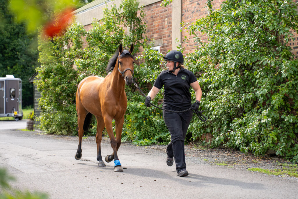
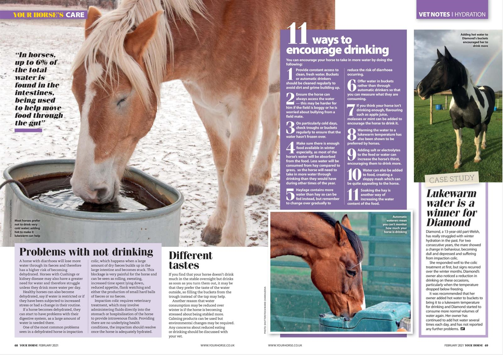
Keeping your horse hydrated
Our Educator Extraordinaire Sarah Hunter shares her knowledge in a fascinating article written for Your Horse Magazine all about the importance of keeping your horse hydrated, what can happen if your horse isn’t well enough hydrated and some advice on how to make sure they’re drinking enough.
Thank you again to Your Horse Magazine for allowing us to share this article.

Lameness investigations
Moving on from estimation in equine lameness investigations
In the last few years, huge strides have been made in this area, making technology for lameness analysis practical and accessible, while also being incredibly reliable and accurate.
This article aims to answer key questions about that technology, to explain why it’s so important and why it should no longer be a vision of the future, but a standard part of the modern lameness examination.
Why measure lameness?
Really, the question should be why not measure lameness. Classic lameness analysis has always been subjective, estimating how lame the horse is and on which leg, on a scale of 1 to 10 or 1 to 5, before trying to decide at which phase in the stride the lameness occurs.
After the treatment or nerve blocks, the vet will try to decide how much the horse has improved through recollection of how much the horse looked before or from their notes.
Wouldn’t it be better if equipment operated by the same professional, who knows how to use it, could measure the exact value of the horse’s lameness and exactly where in the stride the lameness occurred?
Imagine if the same measurement could tell you by what percentage the horse’s lameness has improved after a nerve block or following treatment, so that you know everything is going well. The good news is, this isn’t a pipe dream for the future. That technology is here now!
We were one of the first equine clinics in the UK to adopt objective lameness evaluation
Our RCVS-recognised Advanced Practitioner, Alistair Love has lectured to fellow professionals on objective lameness assessment and how we can use the information to improve treatment from vets, farriers and physiotherapists.
What’s wrong with the traditional method of lameness investigation?
There’s nothing wrong with subjectively evaluating lameness, and we still do it every day on yards. All equine vets are skilled at interpreting lameness this way.
However, recognising lameness is inherently difficult, especially for mild cases; even experienced assessors regularly disagree on the affected limb.
The impact of a resulting misdiagnosis can be profound for both horse and owner. Then there is the case of horses that have variable lameness *sometimes within the same trot-up or sometimes from one trot-up to another), multi-limb lameness or lameness originating from more than one part of the leg!
So, wouldn’t it be better to measure each stride accurately? Then, there’s the placebo effect to consider (where an interpreter is expecting an outcome and so perceives that outcome, even though it isn’t really there).
There are a few other instances in veterinary and human medicine where we estimate a value for something.
We don’t estimate blood pressure or heart rate during an episode of colic or general anaesthesia, and we don’t estimate a tendon injury is improving – so why should lameness be any different?
Can a lameness machine be trusted? Isn’t it risky to rely on the machine?
The fact is we don’t rely on the machine. The machine takes the measurements in the background using Bluetooth, the interpreter is still watching the horse trot-up, looking at every stride, footfall and all the other elements that we use to assess lameness.
We then interpret the results, carefully checking to ensure they are reliable.
The machine is extremely sensitive, but has special confidence intervals built into the results to ensure it has an allowance for normal variation.
It helps to think of it like an x-ray examination. X-rays are only useful if you know how to take them properly, and then you know how to interpret the results.
Objective lameness measurements are no different.
The key, therefore, is being trained to use this equipment. Once you know how to use it and interpret the results, it is extremely valuable.
It’s one important place of the lameness jigsaw that helps you to be certain you’ve put the jigsaw together in the correct way to see the right picture at the end.
Equinosis Q
Equinosis Q – how does it work?
Our RCVS Recognised Advanced Practitioner Alistair Love explains how our modern lameness locator equipment works and how it’s easier to set up and more accessible than you could possibly believe.
The article explains how easy and cost effective it is for everyone to have lameness measurements as part of their horse’s lameness examination, with no added stress for you or your horse.
What equipment does it use?
The system uses three sensors:
- One on the head – to measure vertical head movement.
- One on the pelvis – to measure vertical pelvic movement.
- One of the right forelimb pastern – to tell the machine which leg is on the ground at which time.
- A Bluetooth-connected tablet with lameness locator software.
- A pastern wrap, head bumper and pelvic adhesive pad and clip, to secure the sensors in place.
Did you know?
We were one of the first equine clinics in the UK to adopt objective lameness evaluation.
Our RCVS-recognised Advanced Practitioner, Alistair Love has lectured to fellow professionals on objective lameness assessment and how we can use the information to improve treatment from vets, farriers and physiotherapists.
Does it take long to set up?
Set up is fast and hassle free. The neoprene pastern wrap, neoprene head bumper and pelvic adhesive pad and clip are well tolerated by most horses.
It takes about five minutes to place the sensors and set up the tablet.
Once the sensors are on, they can stay on your horse for the duration of the lameness investigation.
The sensors are extremely robust – in fact, we have firsthand evidence that they can survive a cob rolling on them in the stable!
Is it expensive?
We use the Equinosis-Q lameness locator in virtually every lameness investigation we do.
The best news is that we use it at no additional cost, and charge time for the investigation, plus any additional tests and nerve blocks like we always have.
If we’re performing a re-examination following the initial investigation and treatment, we charge a little bit more to use the equipment to cover the cost of maintaining the equipment and the extra time needed to set it up.
Our clients often request that we use it for re-examinations once they’ve seen how useful and important it is during their horse’s initial work-up.
When used for a re-examination or after a nerve block, the machine can tell you by exactly what percentage the lameness has improved or if it has resolved entirely.
Will It help me to see my horse’s lameness?
Yes! Gone are the days of quietly nodding in agreement with your vet, while you’re unable to see what they’re seeing!
This is the bit most of our owners love the most, as they can see the lameness on the chart, helping both professional and leisure horse owners to understand their horse’s condition better than ever.
We can even email you PDF copies of your horse’s trot-up(s) at your request.
If you have questions about your horse, our experienced team is here to help.
Call us on 01287 623802 or follow us on Facebook (@ClevedaleVetsEquine) for more information.
Equine fact sheets
We’re proud to be part of the VetPartners veterinary group, our respected, like-minded network of vets and vet nurses from around the country.
We’ve put together some information on all the key services that are important for your horse’s health.
Whether you need guidance for current conditions or general support, you’ll find valuable insights to enhance your horse’s wellbeing.